|![[Search]](https://talks.cam.ac.uk/images/search.gif?1209136071) |
|![[A-Z Index]](https://talks.cam.ac.uk/images/az.gif?1209136071) |
|![[Contact]](https://talks.cam.ac.uk/images/contact.gif?1209136071)
| COOKIES: By using this website you agree that we can place Google Analytics Cookies on your device for performance monitoring. | ![[Talks.cam]](https://talks.cam.ac.uk/images/talkslogosmall.gif?1209136071) |
University of Cambridge > Talks.cam > Electron Microscopy Group Seminars > Structure determination by electron crystallography: a powerful complement to conventional methods for structure elucidation
Structure determination by electron crystallography: a powerful complement to conventional methods for structure elucidationAdd to your list(s) Download to your calendar using vCal
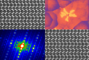
If you have a question about this talk, please contact Duncan Johnstone. Knowledge about the atomic structure of new materials is essential in order to understand its properties, improve its design and develop applications. Modern materials with increasing complexity increases the demands on the structural characterization. Recent developments now makes it possible to obtain 3D single crystal electron diffraction data from crystals down towards the nanometer range. This method has been applied for the structure determination of a large number of new materials, including a significant number of porous materials such as zeolites and Metal-organic Frameworks, where the crystals has been too small for structure determination by conventional methods. Electron microscopy also provides the opportunity of image formation, enabling studies of atomic arrangements at the local scale. This is of importance, especially for materials containing structural disorders. The combination of these two advantages provides unique tools for studies of materials with complex structures. One example is the zeolite ITQ -39, exhibiting a PXRD pattern with a small number of broad peaks. The crystals are needle like, with a cross section of only ~30 nm. By combining 3D electron diffraction data with high resolution transmission electron microscopy (HRTEM) images, the structure of ITQ -39 could be determined.[1] The structure turned out to possess severe disorders including a random intergrowth of three different polytypes and in addition twinning. In a recent study the 3D electron diffraction method was complemented by real space electron tomography at the atomic scale for the structural characterization of a luminescent CuTe. The average atomic structure was determined from the electron diffraction data and subsequently the distribution of Cu vacancies in the material could be studied using electron tomography.[2] References: [1] Willhammar, Zou, Corma et. al., Nature Chem. 4 188 (2012) [2] Willhammar, Liz-Marzán, Bals, van Tendeloo et.al. Nature Comms. 8 14925 (2017) This talk is part of the Electron Microscopy Group Seminars series. This talk is included in these lists:
Note that ex-directory lists are not shown. |
Other listsCambridge Zero Carbon Society Confronting History, the Archive and the 'Stranger' in Educational Research Centre Family Research/Psych Pharmacology Tea Clubs Michaelmas 2014 All CMS events Wall Street meets Lincoln's Inn!Other talksThe formation of high density dust rings and clumps: the role of vorticity Tracking neurobiological factors of language developmental difficulties MEMS Particulate Sensors Future directions panel Perylene-Based Poly(N-Heterocycles): Organic Semiconductors, Biological Fluorescence Probes and Building Blocks for Molecular Surface Networks Insight into the molecular mechanism of extracellular matrix calcification in the vasculature from NMR spectroscopy and electron microscopy |

 Tom Willhammar, Department of Materials and Environmental Chemistry, Stockholm University, Stockholm SE−106 91, Sweden
 Monday 24 April 2017, 11:30-12:30